NV centers in diamond
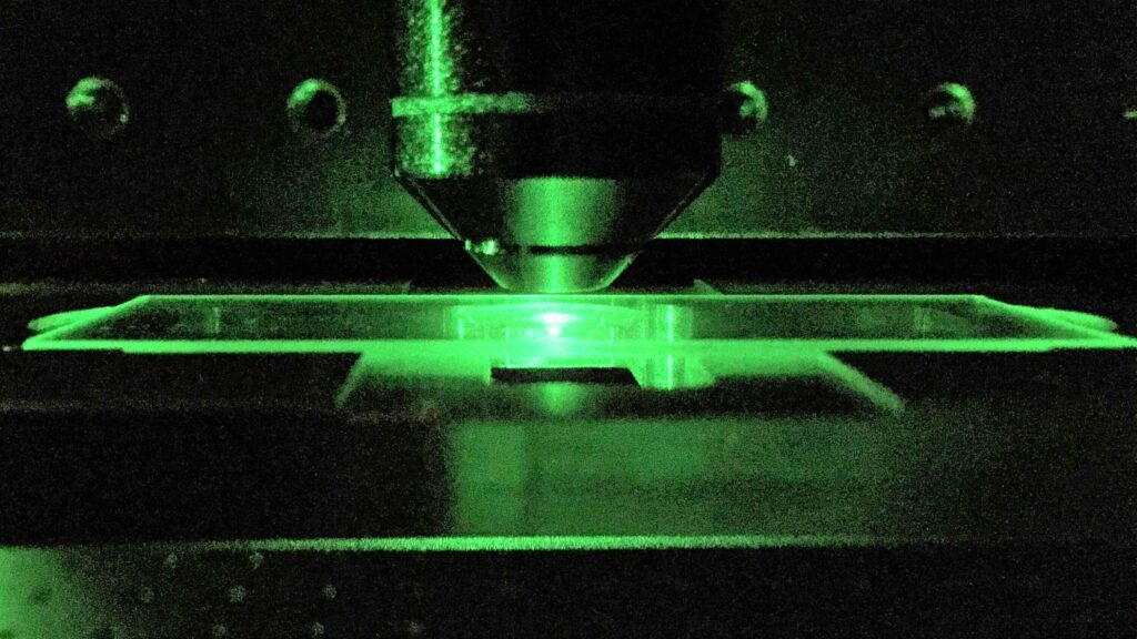
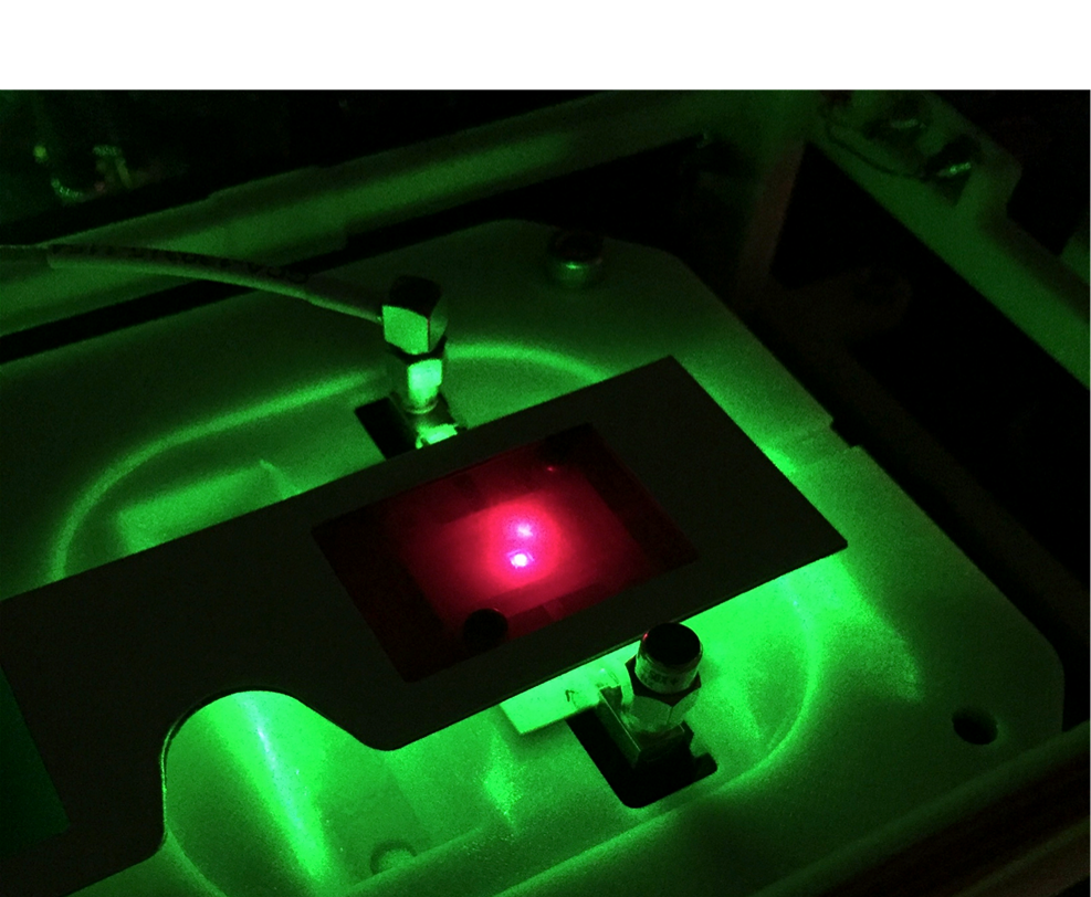
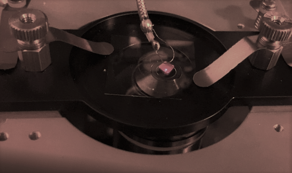
Color centers are defects of the crystal lattice that are capable of absorbing and emitting light. Our group studies the, so called, NV– centers which consist of nitrogen and vacancy at two adjacent locations in the diamond structure (Fig.1). In our research we use diamonds with a concentration of ~ 10 ppm centers. We excite these centers by laser light of with 532 nm wavelength and observe their fluorescence in the range of 600-800 nm. For excitation and detection, confocal microscopy is used which offers excellent spatial resolution.
The ground and the excited electronic states of the NV– center are triplets (S = 1).The non-zero spin of the NV– centres enables their spin polarization. This polarization is created by illuminating the center with light and read out by recording changes in its fluorescence intensity. Polarization changes can be further controlled by microwaves (of about 3 GHz in zero magnetic field Fig.2.) and application of magnetic resonance techniques.

The centers’ properties enable to treat them as artificial atoms in a solid-state matrix. Because of their weak interaction with the diamond lattice, the NV- centers are ideal candidates for experiments in the field of atomic physics, magnetometry and quantum information. Thanks to their strong fluorescence, the NV centers are increasingly often used for imaging and investigating the physical (including magnetic) properties of various nano-scale objects. Moreover, since carbon is a naturally occurring material in biological tissues, NV diamonds are non-toxic and, hence, very attractive for biological studies in vivo.